
Most patients think dental X-rays are only used to detect cavities, but they reveal much more. X-rays help identify bone health concerns, jaw development issues, structural problems, and other conditions that may not be visible during an examination. Detecting these issues early often makes treatment simpler and more effective.
Beyond the Cavity, What X-Rays Are Actually Showing
A full set of dental X-rays at a well-equipped dental practice Wimbledon produces a diagnostic picture that a visual examination simply cannot replicate. The human eye, however experienced the clinician behind it, cannot see through tooth structure, below the gum line, or within the bone of the jaw. X-ray imaging can.
Decay between teeth: Cavities can develop in areas that cannot be seen during a normal examination. X-rays help detect them early, allowing simpler treatment before they become more serious.
Bone health around teeth: Early signs of gum disease and bone loss are often invisible without imaging. Routine X-rays help identify problems before they lead to loose or missing teeth.
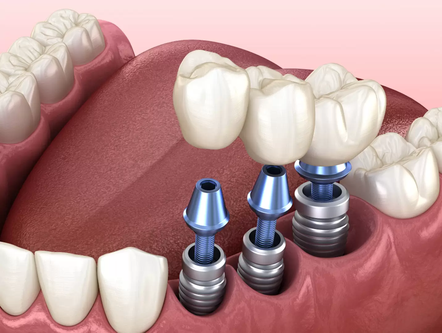
Root structure before treatment: X-rays show the shape and position of tooth roots, helping treatments such as root canals, extractions, and implants to be safer and more predictable.
What Dentists Look for Before Patients Notice Symptoms
One of the reasons routine X-rays are valuable is that dentists are not waiting to find problems once they become painful. Imaging allows patterns of change to be identified while intervention is still simple.
During routine review, clinicians are often assessing for:
- Small changes in enamel density before a cavity forms
- Bone changes around teeth before looseness develops
- Early signs of infection near root tips
- Changes in tooth position and eruption pattern
- Existing restorations are beginning to fail
- Areas of stress or wear that suggest future problems
Most patients leave an appointment feeling nothing has changed. In many cases, that is because imaging allowed the issue to be managed before symptoms ever appeared.
The Early Detection Cases That Change Outcomes Most
Early detection through dental imaging helps identify problems before symptoms appear, often leading to simpler treatment and better long-term outcomes.
Impacted Wisdom Teeth
- Detects positioning issues before pain develops
- Identifies pressure on nearby teeth
- Supports earlier and more predictable treatment
Cysts and Jaw Lesions
- Reveals hidden changes inside the jawbone
- Allows earlier and less invasive intervention
Early Signs of Osteoporosis
- May identify changes in bone density
- Supports earlier medical assessment and ongoing monitoring
Digital X-Ray Technology: Lower Dose, Higher Detail
Modern dental X-ray technology has advanced significantly from traditional film systems. Digital imaging uses substantially lower radiation while producing clearer images that can be enlarged, enhanced, and reviewed over time. For patients concerned about radiation exposure, understanding the comparatively low dose can provide reassurance:
| X-ray type | Radiation dose equivalent |
| Digital dental bitewing X-ray | Natural background radiation for less than a day |
| Full mouth series (digital) | Equivalent to a short domestic flight |
| Panoramic dental X-ray | Equivalent to a few hours of natural background radiation |
The diagnostic information these exposures produce, relative to the dose they deliver, makes routine dental X-ray imaging one of the highest-value clinical tools available in preventive healthcare.
When Dental X-Rays Usually Become More Important
Dental X-rays are not needed at the same frequency for everyone, but they become more valuable when:
- Monitoring developing or erupting teeth
- Reviewing existing dental work
- Assessing changes in gum and bone health
- Investigating unexplained tooth pain
- Planning orthodontic or implant treatment
- Comparing changes after long gaps between visits
The aim is not more X-rays, but using imaging when it provides information that cannot be seen clinically.
Why Routine X-Rays Are an Investment in Avoiding Expensive Treatment
The economics of dental X-rays are straightforward when the alternative is considered. An interproximal cavity found on routine imaging costs a simple composite filling. The same cavity, undetected for two years, may require a root canal and crown, a difference in treatment cost that is multiples of the original filling. A bone lesion found on a routine panoramic X-ray costs a minor surgical procedure. Found two years later, it costs significantly more and carries greater clinical complexity.
Conclusion
Routine X-ray imaging at a dental practice in Wimbledon is not a revenue exercise. It is a clinical discipline that consistently finds the things that a visual examination misses, and finds them at the point where they are still straightforward to treat. Dentist Wimbledon provides comprehensive digital X-ray imaging as a standard component of routine dental care, ensuring that what cannot be seen is examined and that what is found early is treated before it becomes complex.
Author Name: Ankita Patel
Ankita Patel is a dedicated Dentist at The Dental Lounges, located in the heart of Cardiff, UK. With an extensive background in comprehensive patient care and a keen eye for the latest trends in dental health, Ankita serves as a vital resource for both her patients and the broader community. Outside the clinic, she dedicates her time to creating insightful and reader-friendly content for numerous esteemed online platforms.